Friday, 11 December 2020

PLANTAR FASCITIS

PLANTAR FASCITIS (SUBCALCANEAL PAIN)

This is defined as pain on the plantar surface of the heel and is the most common cause of posterior heel pain


Anatomy

The plantar fascia is a long, thin ligament that lies directly beneath the skin on the bottom of your foot. It connects the heel to the front of your foot, and supports the arch of your foot.

Etiopathogensis

Plantar fasciitis has been linked to excessive stress placed on the tissue as a result of athletic activity, muscle weakness or tightness, improper shoes, increase in body weight, aging, inadequate footwear and occupation.3 Plantar fasciitis is usually not the result of a single event but more commonly the result of a history of repetitive micro trauma combined with a biomechanical deficiency of the foot. Finally, degenerative changes that come with age, such as atrophy of the heel fat pad, may also increase ones risk.

Mechanism for development of plantar fasciitis:

Most likely this number is in the thousands. With each
step, the load of the body weight to be applied to the arch causing the arch to drop. This drop in the arch makes the ball of the foot and heel to spread further apart. The fascia in the foot goes into tension to resist this force. If this tension in the fascia is greater than the fascia can handle, the fascia is damaged and the area will become inflamed. The load applied to the foot is divided into two types: Intrinsic load stems from the muscles contracting to move the foot. Much of the intrinsic load applied to the fascia results from the calf muscles. The plantar fascia is part of a larger structure termed the CT band (CT is an acronym for Calf to Toes). The main components of the CT band are the calf, Achilles tendon and plantar fascia. All these components are linked so that tension on any part of the CT band increase tension in the entire system. Of the 3 components of the CT band, the plantar fascia is the weakest link. Extrinsic load refers to all the other loading factors in the plantar fascia other than the intrinsic load. Some of these factors are body weight, frequency of steps, and duration of standing


Clinical Features

The patient complains of pain in the heel, which is more in the morning. It gradually subsides as the patient takes a few steps. The pain increases on prolonged standing, walking, etc.

Clinical Tests

Tenderness can be elicited on the medial aspect of the posterior heel. Passive stretching of the toes increases pain in the heel

Mystifying facts: Why is plantar heel pain more inthe morning?

During sleep, foot is in plantar flexed position causing shortening of the plantar structures. Sudden dorsiflexion in waking up from the night’s sleep stretches the structured abruptly causing pain.

Types of Plantar Fascitis
• Insertional plantar fascitis
-Called the heel pain syndrome
-Pain is felt at the medial calcaneal tubercle
(Point tenderness)

• Diffuse plantar fascitis
-Pain felt diffusely over
the heel and the sole of the foot


Radiographs

Consisting of the routine AP, lateral and oblique views is advised. However, the X-ray does not show any changes in plantar fascitis. It helps to detect calcaneal spur and other heel pathologies.

Treatment

Quick facts: Treatment of plantar fascitis in a nutshell

I line

• NSAIDs.

• Heel pad/cushion.

• Stretching exercises of the ankle and foot.

II line

• Local infiltration of hydrocortisone.

• Custom moulded foot orthosis.

• Soft supportive shoes.

• Foot strapping.

• Stretching exercises.

• Night brace or AFO or short leg walking cast.

III line

Surgery if the entire regime mentioned above fail after one year.
• Measures to reduce pain and inflammation taping,temporary or permanent shoe orthosis, heel cushion, weight management, etc.

• Measures to improve the neurodynamics of the tibial nerve—active calf muscle stretching and calf soft tissue mobilization.

• Joint mobilization with talocalcaneal glides.

• Strengthening the muscles that support the arch namely the posterior tibial, peroneal and intrinsic muscles.

• No response to conservative treatment for three months—LIHC (local infiltration of hydrocorti-sone) is indicated.

• No response to conservative treatment for 6 months—surgery (partial plantar release) is advised.


What is new?

• Endoscopic plantar fasciotomies have a success rate of 85 percent.

• For recalcitrant heel pain instead of surgery 1000 impulses of low energy extracorporeal shock wave treatment (3 applications) is found to be effective.

https://youtu.be/3K7sjNcn3uk

Monday, 2 November 2020

Heart – External Features


Heart-External Features

by physiogyan 


Location and Dimensions  of Heart

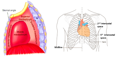
Location: Heart lies in the middle mediastinum. 1/3rd of the heart lies to the right and 2/3rd to the left  of the midline. It lies opposite to T5 - T8 vertebrae in supine position & T6 – T9 vertebrae in erect position.

Dimensions:  Base to apex-12cm;      Transversely- 8-9cm;      Anteroposteriorly- 6cm.

Weight:  In males it weighs: 280-340 gm and in females : 230-280 gm

heart - location

External features of Heart

The heart has:

An apex

  • It is formed by the left ventricle.
  •  It lies in the fifth intercostal space 9 cms. from the median plane.

sternocostal surface of heart

Three surfaces

  • Sternocostal surface: Is formed by right atrium, right ventricle, left auricle and left ventricle.
  • Left surface: Is formed by left auricle and left ventricle.
  • Diaphragmatic surface:  2/3rd is formed by  left ventricle and 1/3rd by right ventricle.
  • Posterior surface/Base: It is quadrilateral in shape.
    • 2/3rd   of base is formed by posterior surface of left atrium and 1/3rd by right atrium.
    •  It lies opposite to T5-T8 vertebra in supine position.

base and diaphragmatic surfaces of heart

Four borders

  • Superior: is formed by the two atria.
  • Right: Right atrium
  • Inferior: Mainly by right ventricle and near the apex by left ventricle.
  • Left: Is formed by left auricle and left ventricle.

Five sulci

  • Atrioventricular sulcus (coronary sulcus): separates atria from ventricles.
  • Anterior interventricular sulcus: separates right and left ventricles on the sternocostal surface.
  • Posterior interventricular sulcus: separates right and left ventricles on the diaphragmatic surface.
  • Interatrial sulcus: Separates right atrium from the left atrium .
  • Sulcus terminalis: extends from the opening of  superior vena cava  to inferior vena cava and separates the rough part from the smooth part of the right atrium.

Applied aspects           

Apex beat: Is the lowermost and outermost thrust of the heart, felt on the front of the chest. In adults it is felt in the left 5th intercostal space 9cm. from the median plane (just medial to the midclavicular line). In infants it is felt in the  3rd intercostal space just lateral to the midclavicular line.

Dextrocardia: It is a congenital anomaly in which the heart lies on the right side of the thoracic cavity. This may be associated with the reversal of all the abdominal organs, a clinical condition known as situs inversus.Dextrocardia


Sunday, 1 November 2020

General Anatomy -Cardiovascular System


Cardiovascular System

by physiogyan 

 General Anatomy -Cardiovascular System


Q. What are the components of the cardiovascular system?
A. Cardiovascular system includes heart and blood vessels i.e. arteries, arterioles, capillaries, venules and veins.


cardiovascular system  
Q. What are the different types of circulation?
A. a. Systemic circulation: It is responsible for transporting oxygenated blood through arteries to the entire body and then returns deoxygenated blood to the heart via veins. The circulation of blood flow is as follows:

systemic circulation


b. Pulmonary circulation: It consist of that part of circulatory system which pumps deoxygenated blood to the lungs and returns oxygenated blood to the heart. The circulation of blood flow is as follows:
  

pulmonary circulation


c. Portal circulation : It is part of systemic circulation. The blood passes through two sets of capillaries,the circulation begins with capillaries and ends with capillaries. The vessel between the two sets of capillaries is known as portal vein. Portal circulation is found at the following sites:
a. Hepatic portal system – between intestines and liver.
b. Renal portal system - in the kidney.
c. Hypothalamo-hypophyseal system – between hypothalamus and hypophysis cerebri.
d. Suprarenal portal system – between the cortex and medulla of adrenal gland.portal circulation
Q. Enumerat the:
a. types of arteries and write examples of each.
b. avascular tissues or structures.
c. structures/organs supplied by end arteries.
d. types of capillaries with examples of each.
e. sites where sinusoids are found.
f. factors responsible for venous return.
g. sites where arterial pulsation is felt.
A. a. Types of arteries:
• Elastic /conducting arteries. e.g. aorta, pulmonary trunk.
• Muscular/distributing arteries . e.g. radial artery, femoral artery etc.elastic and muscular arteries
b. Avascular tissues/structures:
• Epithelium
• Epidermis of skin
• Hair
• Nail
• Cornea
• Cartilage
c. Structures or organs supplied by end arteries:
• Heart
• Kidneys
• Liver
• Brain
• Retina
d. Types of capillaries:
a. Continuous capillaries : e.g. in muscle, brain, connective tissue, skin, lung.
b. Fenestrated capillaries . e.g. in endocrine glands, intestinal villi, renal glomeruli.
continuous and fenstrated capillaries
 
e. Sites where sinusoids are found:
• Liver
• Spleen
• Bone marrow
• Anterior pituitary gland
f. Factors responsible for venous return:
• Contraction of muscles
• Presence of valves in the veins
• Negative intrathoracic pressure
• Pulsation of arteries
g. Sites where arterial pulsations can be felt:
• Carotid artery – along the anterior border of sternocleidomastoid muscle at the level of cricoids cartilage.
• Brachial artery – In front of the elbow medial to the tendon of biceps brachii.
• Radial artery – lateral side of front of forearm at wrist
• Femoral artery – below the inguinal ligament at midinguinal point
• Popliteal artery – in the popliteal fossa
• Dorsalis pedis artery – on the dorsum of foot between the tendon of extensor hallucis longus and extensor digitorum longus.
sites of arterial pulsation
Q. Describe in brief the characteristic features and functions of the arteries,arterioles, capillaries venules and veins.

A .

 
Q. Enumerate the differences between the arteries and veins.
A. Differences between the arteries and veins
Q. Explain the following terms.
a. End arteries
b. Functional arteries
c. Anastomosis
d. Collateral circulation
A. a. End arteries : End arteries are those arteries that do not anastomose with their neighbouring arteries. In case of blockage of an end artery due to a thrombus, the part supplied by it undergoes ischemia and later avascular necrosis. e.g. in kidneys, brain and retina.
b. Functional end arteries: Functional arteries are those arteries whose terminal branches do anastomose, but the anastomosis is not sufficient to maintain the blood supply to the part they supply in case of any blockage in the artery. e.g. coronary arteries.
c. Anastomosis : Anastomosis is defined as communication between the neighbouring blood vessels. It is of two types:
i. Arterial anastomosis : The branches of an artery are connected to the branches of another neighbouring artery. The anastomosis provise collateral channel for circulation when one of the arteries is blocked. e.g. labial branches of facial arteries, intercostals arteries, uterine and ovarian arteries, arterial arcades in the mesentery of intestine etc.
ii. Arteriovenous anastomosis: The direct connection between the arteries and veins without the intervention of capillaries is termed arteriovenous anastomosis. Their function is to regulate temperature and regional blood flow. They are found in the lip, ear nose, nasal mucosa, kidney , intestine etc.
d. Collateral circulation: Collateral circulation is possible when an area of tissue or an organ has a number of different pathways for blood to reach it. This is as a result of anastomosis formed between adjacent blood vessels. In this process small (normally closed) arteries open up and connect two larger arteries or different parts of the same artery and serve alternate route of blood supply.
 

 By physiogyan|  | Tags: anastomosis, avascular sites, collateral circulation, difference between artery and vein, end arteries, important questions on cardiovascular system, types of arteries, types of blood vessels, types of capillaries | Categories: heart


Conducting System and Nerve Supply of Heart

 Conducting System and Nerve Supply of Heart

by physiogyan 

What are the components of conducting system of the heart?

Conducting system of heart is meant for initiating and maintaining cardiac rhythm and establish proper co-ordination between the atrial and ventricular contactions. It is made up of specialized cardiac muscle fibers having a high degree of sensitivity and autorhythmicity.

conducting system of heart

Components of conducting system

Sinuatrial node (SA node)

Is also known as pacemaker.


Initiates the cardiac impulse.


Is located in the upper part of crista terminalis by the side of the opening of superior vena cava.


Impulse from SA node to AV node is carried by intermodal fibers.

triangle of kosch

Atrioventricular node (AV node )

Is situated in the right atrium , in the lower part of interatrial
septum.


It lies in the triangle of Koch, which is bounded by:

Base of septal cusp of tricuspid valve


Orifice of coronary sinus


Tendon of Todaro


 Atrioventricular bundle of HIS

From the AV node it descends in the interventricular septum and divides into:

Right ventricular branch


Left ventricular branch


The two branches descend in the interventricular septum and spread out in the walls of the respective ventricles to end as Purkinje fibers.


Describe briefly the nerve supply of heart.

The heart rate and the cardiac output are controlled by autonomic nervous system.


Sympathetic fibers are provided by the cardiac branches of superior, middle and inferior cervical ganglia (  preganglionic fibers reach from T2-T5 spinal segments).


Parasympathetic fibers are provided by the cardiac branches (superior, inferior and recurrent) of the left & right vagus nerves.


The sympathetic and parasympathetic fibers reach heart via the superficial and deep cardiac plexuses.
Superficial cardiac plexus is located below the arch of aorta. it is formed by:

Cardiac branch of superior cervical ganglion of left sympathetic chain.


Inferior cervical cardiac branch of left vagus.


Deep cardiac plexus is located behind the arch of aorta and in front of tracheal bifurcation. It is formed by:

Cardiac branches of middle and inferior cervical ganglion of both the sympathetic chain and from the superior cervical ganglion of right sympathetic chain.


Cardiac branches of T2-T5 ganglion of both the sympathetic chain.


Superior and recurrent branches of both the vagi and inferior cardiac branch of right vagus.


Physiogyan | | Tags: physiogyan

Friday, 30 October 2020

Venous Drainage of Heartby physiogyan

Venous Drainage of Heart

by physiogyan

 Name the veins that drain heart.

The venous blood from the heart is drained by three system of veins:

Coronary sinus : its tributaries are

great cardiac vein

middle cardiac vein

small cardiac vein

Oblique vein of left atrium

 Anterior cardiac veins

Venae cordis minimae


Describe briefly Coronary sinus and its tributaries.

• Drains most of the venous blood from the heart.
• Is a short , wide venous cannel about 2.5cm. longnand lies in the posterior par of the coronary sulcus.
• Its left end is continuous with the great cardiac vein.
• Its right end opens in the right atrium of the heart between the openings of inferior vena-cava and right atrioventricular orifice.
• Tributaries
Great cardiac vein – lies along anterior interventricular sulcus
Middle cardiac vein –  lies along the posterior interventricular sulcus
Small cardiac vein –lies along  right posterior coronary sulcus
Right marginal vein - along the inferior  boder
Oblique vein of the left atrium
Posterior ventricular vein

venous drainage of heart

Which part of  the heart  is drained by Anterior cardiac veins and where do they open?

• Several small veins, that drain blood from the anterior aspect of right ventricle.
• They open into the right atrium.

What are venae cordis minimae?

• Drain venous blood from the endocardium and deep part of myocardium.
• Open directly into the every chamber of  the heart.

Physiogyan| August 3, 2018 at 7:58 am | Tags: coronary sinus, venous drainage of heart | Categories: Anatomy, Thorax |